Mammography is one of the most widely used methods for breast cancer screening and has contributed significantly to the reduction of the mortality rate through early detection of cancer1. However, the complexity of mammograms and the high volume of exams per radiologist can result in false diagnosis2, 3.

During mammography screening, the presence of breast microcalcifications is a primary risk factor for breast cancer. Breast calcifications in the early stages of breast cancer appear like scattered spots in the mammographic image that range from 0.1 to 1.0 mm in size4. The accurate detection and diagnosis of a breast lesion solely based on mammography findings is difficult and highly depends on the expertise of the radiologist, which leads to a high number of false positives and additional examinations5.

Description

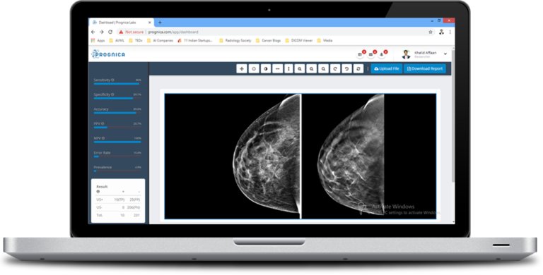
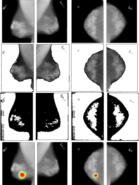
With Prognica’s proprietary “Triple-D Neural Network” technology, PrognicaMMG automatically detects the masses in mammogram and helps in segmentation & classification of the tumor. The process aids in robust diagnosis by using double thresholding to obtain perfect location of tumor. Our neural network strategy uses a smaller number of dense layers with proper feature selection which leads to a higher accuracy in diagnosis of breast cancer, generating location information of detected lesions in the form of heatmaps and abnormality scores reflecting the probability of malignancy.


Training & Validations

  • Trained with a large-scale (>60,000 total cases, >22,000 cancer cases), high-quality DICOM mammogram training dataset.
  • The validation and finalization was made to train the network with 12 features which yields a good training accuracy of 96.1%
  • Clinically validated to significantly improve the interpretive capabilities of radiologists upto ~12%
  • Currently in preparation for CE marking, patent filing

References

1 Li Y, Chen H, Cao L, Ma J. A survey of computer-aided detection of breast cancer with mammography. J Health Med Inf. 2016;4(7).
2 Feig SA. Screening mammography benefit controversies: sorting the evidence. Radiol Clin N Am. 2014;3(52):455–80.
3 Welch HG, Passow HJ. Quantifying the benefits and harms of screening mammography. JAMA Intern Med. 2014;3(174):448–54.
4 Y. Ma, P. C. Tay, R. D. Adams et al., “A novel shape feature to classify microcalcifications”
5 Hamidinekoo, A.; Denton, E.; Rampun, A.; Honnor, K.; Zwiggelaar, R. Deep learning in mammography and breast histology, [CrossRef] [PubMed]